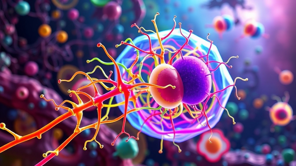
The cytoskeleton is a complex and dynamic network of protein filaments that extends throughout the cytoplasm of eukaryotic cells. Much like the skeleton in the human body, the cytoskeleton provides structural support, but it also plays a far more diverse set of roles. It maintains the cell’s shape, facilitates movement, enables intracellular transport, and coordinates many of the biochemical activities necessary for life. The cytoskeleton is not a static structure but a highly adaptable system that responds to the needs of the cell, constantly assembling and disassembling to support a variety of functions.

In this article, we will explore the structure and function of the cytoskeleton, focusing on its three primary components—microfilaments, intermediate filaments, and microtubules—and their roles in maintaining cell integrity and facilitating crucial biological processes. By understanding the cytoskeleton’s importance, we can appreciate its role in everything from cell division and migration to the transport of materials within cells.
Structure of the Cytoskeleton
The cytoskeleton is composed of three types of protein filaments, each with distinct structural and functional characteristics: microfilaments (also known as actin filaments), intermediate filaments, and microtubules. These components work together to provide mechanical support, facilitate cellular movement, and enable the transport of materials within the cell.
1. Microfilaments (Actin Filaments)
Microfilaments, also called actin filaments, are the thinnest filaments of the cytoskeleton, with a diameter of approximately 7 nanometers. They are composed of the protein actin, which forms long, thin fibers arranged in a double helical structure. These filaments are highly dynamic, meaning they can rapidly assemble and disassemble in response to the cell’s needs.
Functions of Microfilaments
- Cell Shape and Structural Support:
Microfilaments are key to maintaining the cell’s shape, especially in animal cells that lack cell walls. They form a dense network beneath the plasma membrane, providing structural support and resisting deformation.Example: In red blood cells, the actin cytoskeleton helps maintain their characteristic biconcave shape, which is essential for efficient oxygen transport. - Cell Movement and Motility:
Microfilaments are involved in cell motility through processes like cell crawling and cytoplasmic streaming. Actin filaments, along with myosin (a motor protein), enable muscle contraction and play a critical role in the movement of non-muscle cells, such as amoeboid movement in white blood cells.Example: In immune cells like macrophages, actin filaments drive cell migration, enabling the cell to move toward infection sites in a process known as chemotaxis. - Cytokinesis:
During cell division, microfilaments form the contractile ring that pinches the cell in two during cytokinesis (the final stage of mitosis or meiosis). The ring contracts, dividing the cytoplasm and creating two daughter cells.Example: In animal cells, actin filaments assemble into a contractile ring at the equator of the dividing cell during cytokinesis, ensuring the proper separation of the two new cells.
2. Intermediate Filaments
Intermediate filaments are named for their diameter, which is about 10 nanometers, placing them between microfilaments and microtubules in terms of size. These filaments are composed of a variety of proteins, depending on the cell type, and are more stable than microfilaments and microtubules. Their primary function is to provide mechanical strength to cells.
Functions of Intermediate Filaments
- Structural Integrity and Resilience:
Intermediate filaments are crucial for maintaining the cell’s mechanical integrity. They form a durable scaffold that helps the cell withstand mechanical stress, such as stretching or compression, without rupturing.Example: Keratin is a type of intermediate filament found in epithelial cells. It is particularly abundant in skin, hair, and nails, where it provides strength and protection. In the skin, keratin filaments help cells resist abrasion and tearing, ensuring the integrity of the outermost layers of the body. - Anchoring Organelles:
Intermediate filaments help anchor organelles like the nucleus in place within the cell. This organization is essential for maintaining cellular structure and ensuring the proper distribution of organelles during cell division.Example: Lamin proteins, a type of intermediate filament, form a supportive meshwork around the inside of the nuclear envelope, maintaining the shape and stability of the nucleus. - Cell-Cell Junctions:
In tissues, intermediate filaments provide structural support by linking to cell-cell junctions, such as desmosomes, which are points of contact between adjacent cells. This network helps distribute mechanical stress across the tissue.Example: In the heart and skin, intermediate filaments connect cells at desmosomes, allowing tissues to maintain cohesion and resist mechanical stress, particularly in tissues that experience frequent stretching and contraction.
3. Microtubules
Microtubules are the largest filaments in the cytoskeleton, with a diameter of about 25 nanometers. They are composed of tubulin proteins, which form hollow, tube-like structures. Microtubules are highly dynamic, constantly growing and shrinking as tubulin subunits are added or removed.
Functions of Microtubules
- Intracellular Transport:
One of the primary roles of microtubules is to act as tracks for the transport of organelles, vesicles, and proteins within the cell. Motor proteins, such as kinesin and dynein, “walk” along microtubules, carrying cargo to different parts of the cell.Example: In neurons, microtubules play a crucial role in transporting neurotransmitters from the cell body to the axon terminal, ensuring proper communication between nerve cells. - Cell Division (Mitotic Spindle Formation):
During cell division, microtubules form the mitotic spindle, a structure that separates chromosomes into the daughter cells. The spindle fibers attach to the centromeres of chromosomes and pull them apart during anaphase of mitosis and meiosis.Example: In human cells, the mitotic spindle ensures that each daughter cell receives an identical set of chromosomes during mitosis, maintaining genetic stability across generations of cells. - Cell Movement (Cilia and Flagella):
Microtubules are the structural components of cilia and flagella, which are specialized for movement. Cilia are short, hair-like projections that move fluid over the surface of the cell, while flagella are longer and whip-like, enabling cell motility.Example: In the respiratory tract, cilia beat in a coordinated fashion to move mucus and trapped particles out of the lungs, helping to protect the respiratory system from infection.
Dynamic Nature of the Cytoskeleton
One of the most remarkable features of the cytoskeleton is its ability to rapidly change in response to the needs of the cell. This dynamic instability is particularly evident in microfilaments and microtubules, which can grow or shrink depending on cellular signals. The ability to quickly assemble or disassemble allows the cytoskeleton to adapt to changes in the environment or to perform different cellular functions as needed.
Polymerization and Depolymerization
Both microfilaments and microtubules undergo polymerization (the process of adding subunits to grow the filament) and depolymerization (the process of removing subunits to shrink the filament). This cycle allows the cytoskeleton to be flexible and responsive.
Example: During cell migration, actin filaments in the leading edge of the cell polymerize to push the plasma membrane forward, while filaments at the rear depolymerize to allow the cell to contract and move.
The Role of the Cytoskeleton in Disease
Given its critical role in maintaining cellular function, it is no surprise that defects in the cytoskeleton can lead to a variety of diseases. Mutations in cytoskeletal proteins can disrupt normal cell function, leading to conditions that affect movement, division, and structural integrity.
Cancer
In cancer, abnormalities in the cytoskeleton can contribute to uncontrolled cell division and metastasis. Microtubule-targeting drugs, such as taxanes (e.g., paclitaxel), are used in chemotherapy to disrupt the mitotic spindle, preventing cancer cells from dividing.
Example: Paclitaxel, a chemotherapy drug, stabilizes microtubules, preventing their depolymerization. This disrupts the ability of cancer cells to undergo mitosis, leading to cell death.
Neurodegenerative Diseases
In neurodegenerative diseases like Alzheimer’s and Parkinson’s, cytoskeletal proteins become abnormally regulated, leading to the collapse of cellular transport systems. Disruption in the microtubule network can impair the transport of essential molecules within neurons, contributing to cell death.
Example: In Alzheimer’s disease, the protein tau, which normally stabilizes microtubules, becomes hyperphosphorylated and forms tangles, disrupting the microtubule network and leading to neuronal dysfunction and degeneration.
Muscular Dystrophy
Mutations in cytoskeletal components, such as dystrophin, can lead to diseases like Duchenne muscular dystrophy. Dystrophin is a protein that links the cytoskeleton of muscle cells to the extracellular matrix, providing structural support. In its absence, muscle cells become damaged and progressively weaken over time.
Example: In patients with Duchenne muscular dystrophy, the lack of functional dystrophin leads to the breakdown of muscle tissue, resulting in muscle weakness and eventual loss of mobility.
Conclusion
The cytoskeleton is an essential and versatile structure that not only provides the cell with mechanical strength but also facilitates intracellular transport, cell division, and movement. Comprising microfilaments, intermediate filaments, and microtubules, the cytoskeleton forms a dynamic scaffold that is continuously remodeled to meet the needs of the cell. Its importance in various cellular processes, from maintaining structural integrity to enabling cell migration, highlights the complexity and adaptability of life at the microscopic level.
By understanding the cytoskeleton and its components, we gain valuable insights into how cells function in both health and disease. Whether driving cell movement, transporting vital molecules, or ensuring the faithful segregation of chromosomes, the cytoskeleton is truly the framework that sustains the cellular world.