A eukaryotic cell is a type of cell that has a defined nucleus and various other membrane-bound organelles. Eukaryotic cells are more complex than prokaryotic cells (such as bacteria), which lack a nucleus and most organelles. Organisms composed of eukaryotic cells include animals, plants, fungi, and protists, collectively known as eukaryotes. Eukaryotic cells perform a wide range of functions, from energy production to reproduction, allowing multicellular organisms to grow, develop, and maintain homeostasis.
In this article, we will explore the structure of eukaryotic cells, their functions, and their role in life. By examining the different organelles and their roles, along with examples of eukaryotic organisms, we will gain a comprehensive understanding of how these cells sustain complex life forms.
Key Characteristics of Eukaryotic Cells
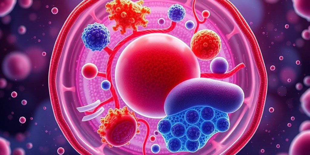
Eukaryotic cells are distinguished by their membrane-bound nucleus and the presence of other membrane-bound organelles, such as mitochondria, endoplasmic reticulum, and Golgi apparatus. These organelles compartmentalize the cell’s functions, allowing specific processes to occur in designated regions, making eukaryotic cells highly efficient and specialized.
General Characteristics of Eukaryotic Cells:
- Membrane-Bound Nucleus: Eukaryotic cells have a nucleus that contains the cell’s genetic material (DNA) and regulates gene expression. The nuclear envelope, a double membrane, separates the nucleus from the cytoplasm.
- Organelles: Eukaryotic cells have a variety of organelles that carry out specific functions, such as energy production (mitochondria), protein synthesis (ribosomes), and waste disposal (lysosomes).
- Cytoskeleton: A network of protein fibers, including microtubules, actin filaments, and intermediate filaments, that provides structural support, helps in cell division, and facilitates the movement of organelles within the cell.
- Cell Size: Eukaryotic cells are generally larger than prokaryotic cells, ranging from 10 to 100 micrometers in diameter.
- Multicellularity: Many eukaryotic organisms, such as animals, plants, and fungi, are multicellular, with specialized cell types that perform different functions. Some eukaryotes, like amoebas and yeast, are unicellular.
Structure of Eukaryotic Cells
Eukaryotic cells have a variety of organelles, each with a specific function that contributes to the cell’s overall operation. These organelles work together to ensure the cell can grow, divide, produce energy, and synthesize essential molecules.
1. Nucleus: The Control Center
The nucleus is the defining feature of eukaryotic cells. It houses the cell’s genetic material (DNA) in the form of chromatin, which is organized into chromosomes during cell division. The nucleus is surrounded by a nuclear envelope, a double membrane that separates the nuclear contents from the cytoplasm. Nuclear pores in the envelope allow the exchange of materials between the nucleus and cytoplasm, including RNA and ribosomal subunits.
The nucleolus, a dense region within the nucleus, is responsible for synthesizing ribosomal RNA (rRNA) and assembling ribosomes. Ribosomes, which are later transported to the cytoplasm, are essential for protein synthesis.
Function of the Nucleus:
- DNA Storage: The nucleus stores the genetic information required for the growth, development, and functioning of the cell.
- Gene Regulation: The nucleus controls gene expression, determining which proteins are synthesized and when, allowing the cell to respond to environmental signals.
Example: Nucleus in Muscle Cells
In skeletal muscle cells, multiple nuclei are present within each cell. This multinucleation allows the muscle cells to produce large amounts of proteins required for muscle contraction and repair, supporting the heavy workload these cells must perform.
2. Mitochondria: The Powerhouse of the Cell
Mitochondria are often referred to as the “powerhouses” of the cell because they generate most of the cell’s adenosine triphosphate (ATP), the energy currency of the cell. Mitochondria have a double membrane, with an inner membrane folded into structures called cristae, which increase the surface area for ATP production. Inside the mitochondrion is the matrix, which contains enzymes involved in the Krebs cycle and oxidative phosphorylation.
Mitochondria have their own DNA and ribosomes, which resemble those of prokaryotes, supporting the theory that they originated from an ancient symbiotic relationship between a primitive eukaryotic cell and a prokaryotic organism.
Function of Mitochondria:
- ATP Production: Mitochondria produce ATP through cellular respiration, a process that converts nutrients into usable energy.
- Apoptosis: Mitochondria play a role in programmed cell death, or apoptosis, a process essential for removing damaged or unnecessary cells.
Example: Mitochondria in Muscle Cells
In cardiac muscle cells, mitochondria are abundant due to the high energy demands of the heart. The constant contraction and relaxation of heart muscles require large amounts of ATP, which is generated by the mitochondria.
3. Endoplasmic Reticulum (ER): Protein and Lipid Synthesis
The endoplasmic reticulum (ER) is a network of membranes that extends throughout the cytoplasm. There are two types of ER: rough ER (RER) and smooth ER (SER), each with different functions.
- Rough ER: The rough ER is studded with ribosomes, giving it a “rough” appearance. It is involved in the synthesis of proteins, particularly those that are destined for secretion or for use in the cell membrane.
- Smooth ER: The smooth ER lacks ribosomes and is involved in the synthesis of lipids, metabolism of carbohydrates, and detoxification of drugs and poisons. It also plays a role in the storage of calcium ions, which are important in muscle contraction.
Function of ER:
- Protein Synthesis (Rough ER): The rough ER is responsible for producing proteins that are either secreted from the cell or inserted into cellular membranes.
- Lipid Metabolism (Smooth ER): The smooth ER synthesizes lipids, such as phospholipids and cholesterol, which are essential components of cellular membranes.
Example: ER in Liver Cells
In liver cells, the smooth ER is highly developed to facilitate the detoxification of harmful substances, such as alcohol and drugs, by breaking them down into less toxic forms.
4. Golgi Apparatus: Packaging and Transport
The Golgi apparatus is a series of flattened, membrane-bound sacs known as cisternae. It acts as the cell’s “post office,” modifying, sorting, and packaging proteins and lipids that are synthesized in the ER. These molecules are then transported in vesicles to their final destinations, either within the cell or outside of it through exocytosis.
Function of Golgi Apparatus:
- Protein Modification: The Golgi apparatus modifies proteins by adding carbohydrate or lipid groups to them, creating glycoproteins and lipoproteins.
- Vesicle Transport: The Golgi packages these molecules into vesicles for transport to different parts of the cell or for secretion outside the cell.
Example: Golgi Apparatus in Gland Cells
In pancreatic cells, the Golgi apparatus plays a critical role in processing and packaging digestive enzymes that are then secreted into the small intestine to aid in digestion.
5. Lysosomes: The Cell’s Digestive System
Lysosomes are membrane-bound organelles that contain hydrolytic enzymes responsible for breaking down cellular waste, damaged organelles, and invading pathogens. Lysosomes function as the cell’s “recycling centers,” digesting macromolecules into their basic building blocks, which can be reused by the cell.
Function of Lysosomes:
- Intracellular Digestion: Lysosomes digest cellular debris and foreign particles, such as bacteria and viruses.
- Autophagy: Lysosomes participate in autophagy, a process where the cell breaks down its own damaged organelles to maintain cellular health.
Example: Lysosomes in Immune Cells
In macrophages (a type of immune cell), lysosomes digest bacteria and viruses that have been engulfed through a process known as phagocytosis. This process helps the body defend against infections.
6. Cytoskeleton: Cell Shape and Movement
The cytoskeleton is a network of protein fibers that provides structural support for the cell, helps maintain its shape, and aids in movement. The cytoskeleton is composed of three types of protein filaments: microtubules, actin filaments, and intermediate filaments.
- Microtubules: Hollow tubes made of tubulin that serve as tracks for the movement of organelles and vesicles. Microtubules are also involved in cell division and the formation of the mitotic spindle.
- Actin Filaments (Microfilaments): Thin fibers that play a role in cell movement, muscle contraction, and cell division.
- Intermediate Filaments: Provide structural stability to the cell and help anchor organelles in place.
Function of the Cytoskeleton:
- Structural Support: The cytoskeleton provides the mechanical strength needed to maintain the cell’s shape.
- Cell Movement: The cytoskeleton facilitates cell motility through structures like cilia, flagella, and pseudopodia.
- Cell Division: Microtubules form the mitotic spindle, which helps segregate chromosomes during cell division.
Example: Cytoskeleton in Nerve Cells
In neurons, microtubules help transport neurotransmitters and other molecules along the length of the axon, allowing signals to be transmitted from one part of the neuron to another.
7. Vacuoles: Storage and Regulation
Vacuoles are large, membrane-bound sacs found in both plant and animal cells, although they are much larger and more prominent in plant cells. In plants, vacuoles store water, nutrients, and waste products. They also help maintain turgor pressure, which keeps the plant rigid and upright.
Function of Vacuoles:
- Storage: Vacuoles store essential nutrients, water, and waste products.
- Waste Disposal: Vacuoles help isolate harmful materials and facilitate their breakdown.
- Turgor Pressure: In plant cells, vacuoles maintain turgor pressure, which is essential for structural support.
Example: Vacuoles in Plant Cells
In plant cells, the central vacuole occupies a large portion of the cell’s volume and is filled with water and dissolved substances. When the vacuole is full, it pushes against the cell wall, providing structural support to the plant.
8. Chloroplasts: Photosynthesis in Plant Cells
Chloroplasts are specialized organelles found in plant cells and some protists. They are responsible for photosynthesis, the process by which light energy is converted into chemical energy in the form of glucose. Chloroplasts contain the green pigment chlorophyll, which captures light energy.
Chloroplasts have a double membrane and contain their own DNA, much like mitochondria. Inside the chloroplasts are thylakoid membranes, which are organized into stacks called grana. The light-dependent reactions of photosynthesis occur in these membranes, while the Calvin cycle takes place in the stroma, the fluid-filled space inside the chloroplast.
Function of Chloroplasts:
- Photosynthesis: Chloroplasts convert sunlight into glucose, which serves as an energy source for the plant.
- Oxygen Production: Chloroplasts release oxygen as a byproduct of photosynthesis.
Example: Chloroplasts in Leaf Cells
In leaf cells of green plants, chloroplasts are abundant, capturing sunlight to produce energy for the plant. This is particularly evident in plants like spinach, where chloroplasts are densely packed within the cells to maximize photosynthesis.
Eukaryotic Cells in Multicellular Organisms
In multicellular organisms, eukaryotic cells often specialize to perform specific functions. This specialization, known as cell differentiation, allows organisms to develop complex structures, such as tissues and organs, each composed of distinct cell types that work together to maintain homeostasis.
For example, in animals, muscle cells are specialized for contraction, nerve cells for signal transmission, and red blood cells for oxygen transport. In plants, leaf cells are optimized for photosynthesis, while root cells absorb water and nutrients.
Example: Eukaryotic Cells in the Human Body
The human body contains a wide variety of specialized eukaryotic cells:
- Epithelial cells line the surfaces of organs and act as barriers against pathogens.
- Neurons transmit electrical signals throughout the nervous system.
- Osteocytes are bone cells that maintain the strength and structure of bones.
Conclusion
Eukaryotic cells are the building blocks of complex life forms, characterized by their membrane-bound organelles, including the nucleus, mitochondria, endoplasmic reticulum, and Golgi apparatus. These cells carry out essential functions such as energy production, protein synthesis, waste disposal, and cellular communication. Their ability to compartmentalize these functions into distinct organelles allows eukaryotic cells to support the growth and development of multicellular organisms, from simple protists to highly complex plants and animals.
Understanding the structure and function of eukaryotic cells is fundamental to biology, as these cells form the basis for all complex life on Earth. Their versatility, efficiency, and specialization allow organisms to perform the vast array of tasks required for survival, adaptation, and reproduction.