The human heart is a muscular organ at the center of the circulatory system, responsible for pumping blood throughout the body. It plays an indispensable role in delivering oxygen and nutrients to tissues, removing waste products like carbon dioxide, and maintaining the overall homeostasis of the body. Though it weighs only about 250-350 grams and is roughly the size of a fist, the heart is vital for sustaining life.
In this article, we will explore the anatomy of the heart, its functions, how it maintains circulation, and its significance in overall human health. Through examples of heart function in daily life, as well as common heart conditions, we will illustrate why the heart is one of the most crucial organs in the human body.
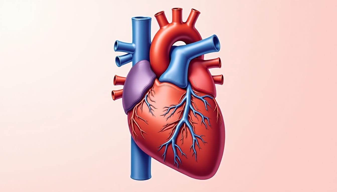
Anatomy of the Human Heart
The human heart is located in the thoracic cavity, between the lungs, slightly to the left of the midline. It is enclosed in a protective sac called the pericardium, which helps to anchor the heart in place while allowing it to expand and contract. The heart is composed of four chambers—two upper chambers called atria and two lower chambers called ventricles—and is divided into left and right sides by a muscular wall known as the septum.
Chambers of the Heart
- Atria: The atria are the upper chambers of the heart that receive blood. The right atrium receives deoxygenated blood from the body through the superior and inferior vena cava, while the left atrium receives oxygenated blood from the lungs via the pulmonary veins.
- Ventricles: The ventricles are the lower chambers that pump blood out of the heart. The right ventricle pumps deoxygenated blood to the lungs through the pulmonary artery for oxygenation, and the left ventricle pumps oxygenated blood to the rest of the body via the aorta. The left ventricle is the most powerful chamber, as it needs to generate enough force to circulate blood throughout the entire body.
Valves of the Heart
The heart contains four valves that ensure blood flows in one direction, preventing backflow:
- Tricuspid Valve: Located between the right atrium and right ventricle, the tricuspid valve prevents blood from flowing back into the right atrium when the right ventricle contracts.
- Pulmonary Valve: This valve is located between the right ventricle and the pulmonary artery. It ensures that blood flows from the right ventricle to the lungs without regurgitating back into the ventricle.
- Mitral Valve: Also known as the bicuspid valve, the mitral valve is between the left atrium and left ventricle. It prevents backflow of blood into the left atrium during the powerful contractions of the left ventricle.
- Aortic Valve: Positioned between the left ventricle and the aorta, the aortic valve allows blood to flow from the heart to the rest of the body but prevents it from returning to the left ventricle.
Layers of the Heart Wall
The heart wall is composed of three layers, each with distinct functions:
- Epicardium: This is the outermost layer of the heart wall, made up of connective tissue. It serves as a protective layer and also contains blood vessels that nourish the heart.
- Myocardium: The middle layer and the thickest part of the heart wall, the myocardium is composed of cardiac muscle tissue. This is the layer responsible for the heart’s pumping action. The myocardium’s contraction is what drives blood out of the heart and into circulation.
- Endocardium: The innermost layer, the endocardium lines the inside of the heart chambers and covers the heart valves. It ensures smooth blood flow within the heart and plays a role in regulating the function of the heart valves.
How the Heart Functions: The Cardiac Cycle
The heart functions through a continuous cycle of contraction and relaxation known as the cardiac cycle, which is divided into two main phases: systole and diastole.
- Systole: During systole, the ventricles contract, pumping blood out of the heart. The right ventricle sends blood to the lungs via the pulmonary artery, while the left ventricle sends blood to the body through the aorta. At the same time, the atrioventricular valves (tricuspid and mitral) close to prevent backflow into the atria.
- Diastole: In diastole, the heart relaxes, allowing the atria to fill with blood. The ventricles also relax and expand to receive blood from the atria. The semilunar valves (pulmonary and aortic) close to prevent blood from flowing back into the ventricles from the arteries.
The cardiac cycle ensures that the heart pumps blood efficiently, maintaining a continuous flow of oxygenated and deoxygenated blood to and from the lungs and the rest of the body.
Electrical Conduction System of the Heart
The heart has an intrinsic electrical conduction system that regulates its rhythmic contractions. This system ensures that the atria and ventricles contract in a coordinated manner, optimizing blood flow. The key components of this system include:
- Sinoatrial (SA) Node: Often referred to as the heart’s natural pacemaker, the SA node is located in the right atrium and generates electrical impulses that initiate each heartbeat. These impulses cause the atria to contract, pushing blood into the ventricles.
- Atrioventricular (AV) Node: Located between the atria and ventricles, the AV node receives the electrical impulse from the SA node and delays it slightly before passing it on to the ventricles. This delay allows the ventricles to fill with blood before they contract.
- Bundle of His and Purkinje Fibers: After passing through the AV node, the electrical signal travels down the Bundle of His and into the Purkinje fibers, which distribute the impulse through the ventricles, triggering their contraction.
This electrical conduction system enables the heart to maintain a regular and synchronized heartbeat, typically between 60 and 100 beats per minute in a healthy adult at rest.
Blood Circulation: Systemic and Pulmonary Circuits
The heart plays a central role in the circulatory system, which consists of two main circuits: the systemic circuit and the pulmonary circuit.
1. Pulmonary Circulation
Pulmonary circulation is the path of blood between the heart and the lungs. Its primary function is to oxygenate the blood and remove carbon dioxide. The process works as follows:
- Deoxygenated blood from the body returns to the right atrium through the superior and inferior vena cava.
- This blood is pumped into the right ventricle, which then sends it to the lungs through the pulmonary artery.
- In the lungs, the blood releases carbon dioxide and picks up oxygen.
- Oxygenated blood then returns to the left atrium of the heart via the pulmonary veins.
2. Systemic Circulation
Systemic circulation is the process by which oxygenated blood is distributed to the tissues of the body. After the blood is oxygenated in the lungs, it follows this pathway:
- The oxygen-rich blood enters the left atrium and is pumped into the left ventricle.
- The left ventricle then contracts, sending the blood through the aorta to the rest of the body.
- As the blood travels through the body’s tissues, it delivers oxygen and nutrients while collecting carbon dioxide and other waste products.
- Deoxygenated blood then returns to the right atrium, completing the cycle.
Together, pulmonary and systemic circulation ensure that the body’s cells receive the oxygen and nutrients they need to function, while also removing waste products.
Importance of the Human Heart in Health
The heart is central to human health, and maintaining its function is critical for survival. Any dysfunction in the heart can lead to serious, life-threatening conditions. Here are some key examples of heart-related health issues:
1. Coronary Artery Disease (CAD)
Coronary artery disease is one of the leading causes of death globally. It occurs when the coronary arteries, which supply blood to the heart muscle, become narrowed or blocked due to the buildup of plaque (atherosclerosis). Reduced blood flow to the heart can cause chest pain (angina) or lead to a heart attack.
Example: A person with coronary artery disease may experience shortness of breath or chest discomfort, especially during physical activity. If left untreated, a complete blockage of the coronary arteries can lead to a myocardial infarction (heart attack), where part of the heart muscle dies due to lack of oxygen.
2. Heart Failure
Heart failure occurs when the heart is unable to pump blood effectively, leading to insufficient blood flow to meet the body’s needs. This can be caused by various factors, including weakened heart muscle (cardiomyopathy), high blood pressure, or previous heart attacks that have damaged the heart.
Example: A person with heart failure may experience fatigue, swelling in the legs (edema), and difficulty breathing, particularly when lying down. Heart failure can significantly reduce a person’s quality of life and, if severe, can be fatal.
3. Arrhythmias
Arrhythmias are irregular heartbeats caused by disturbances in the heart’s electrical conduction system. These can range from harmless irregularities to life-threatening conditions like ventricular fibrillation, where the heart quivers instead of pumping blood, leading to sudden cardiac arrest.
Example: A person with an arrhythmia such as atrial fibrillation may feel palpitations, dizziness, or fatigue. While some arrhythmias are benign, others may require medical intervention, such as medications or the implantation of a pacemaker.
4. Hypertension (High Blood Pressure)
Hypertension is a condition where the force of the blood against the artery walls is consistently too high. Over time, this can strain the heart, leading to complications like heart attack, stroke, or heart failure.
Example: While hypertension often has no symptoms, it silently damages the arteries and increases the risk of cardiovascular events. Regular monitoring and lifestyle changes, such as reducing salt intake and exercising, can help manage hypertension.
Conclusion
The human heart is an extraordinary organ that serves as the engine of life, driving the circulation of blood throughout the body. Its intricate structure—comprising chambers, valves, and a powerful electrical system—ensures that oxygenated blood reaches tissues while deoxygenated blood is directed to the lungs for re-oxygenation.
Understanding the heart’s anatomy, function, and role in circulation is key to recognizing how vital it is for overall health. The heart’s ability to pump blood continuously throughout our lives is a testament to its strength and resilience. However, conditions like coronary artery disease, heart failure, arrhythmias, and hypertension remind us that the heart’s health must be maintained through a healthy lifestyle, regular check-ups, and medical intervention when necessary.
By appreciating the complexity and importance of the human heart, we gain insight into the central role it plays in keeping us alive and healthy.